What does the body of an amoeba consist of? Common amoeba
Read also
Amoeba is a representative of single-celled animals capable of actively moving with the help of special specialized organelles. The structural features and significance of these organisms in nature will be revealed in our article.
Characteristics of the subkingdom Protozoa
Despite the fact that protozoa have this name, their structure is quite complex. After all, one microscopic cell is capable of performing the functions of an entire organism. Amoeba is another proof that an organism up to 0.5 mm in size is capable of breathing, moving, reproducing, growing and developing.
Protozoan movement
Single-celled organisms move with the help of special organelles. In ciliates they are called cilia. Just imagine: on the surface of a cell, up to 0.3 mm in size, there are about 15 thousand of these organelles. Each of them makes pendulum-like movements.
Euglena has a flagellum. Unlike cilia, it makes helical movements. But what these organelles have in common is that they are permanent outgrowths of the cell.
The movement of the amoeba is due to the presence of pseudopods. They are also called pseudopodia. These are unstable cellular structures. Due to the elasticity of the membrane, they can form anywhere. First, the cytoplasm moves outward and a protrusion is formed. Then the reverse process follows, the pseudopods are directed into the cell. As a result, the amoeba moves slowly. The presence of pseudopods is distinctive characteristic feature this representative of the subkingdom Unicellular.

Amoeba proteus

Amoeba structure
All protozoan cells are eukaryotic - they contain a nucleus. The organs of the amoeba, or rather its organelles, are capable of carrying out all life processes. The pseudopods are not only involved in movement, but also provide the amoeba with nutrition. With their help, a single-celled animal embraces a food particle, which is surrounded by a membrane and ends up inside the cell. This is the process of formation of digestive vacuoles in which the breakdown of substances occurs. This method of absorption of solid particles is called phagocytosis. Undigested food remains are released anywhere in the cell through the membrane.

Amoeba, like all protozoa, does not have specialized respiratory organelles, carrying out gas exchange through the membrane.
But the process of regulation of intracellular pressure is carried out with the help of contractile vacuoles. The salt content in the environment is higher than inside the body itself. Therefore, according to the laws of physics, water will flow into the amoeba - from an area with a higher concentration to a lower one. regulate this process by removing some metabolic products along with water.
Amoebas are characterized by asexual reproduction by two. This is the most primitive of all known methods, however, it provides accurate storage and transmission hereditary information. In this case, first the organelles occur, and then the separation of the cell membrane occurs.
This simplest organism able to respond to factors environment: light, temperature, change chemical composition reservoir
Single-celled organisms tolerate unfavorable conditions in the form of cysts. Such a cell stops moving, its water content decreases, and the pseudopods are retracted. And it itself is covered with a very dense shell. This is a cyst. When advancing favorable conditions The amoebas emerge from the cysts and proceed to normal life processes.
Dysenteric amoeba
Many species of these protozoa also play a positive role in nature. Amoebas are a source of food for many animals, namely fry of fish, worms, mollusks, and small crustaceans. They clean fresh water bodies of bacteria and rotting algae and are an indicator of the cleanliness of the environment. took part in the formation of limestone and chalk deposits.
This class includes single-celled animals that are characterized by a variable body shape. This is due to the formation of pseudopods, which serve to move and capture food. Many rhizomes have an internal or external skeleton in the form of shells. After death, these skeletons settle to the bottom of reservoirs and form silt, which gradually turns into chalk.
A typical representative of this class is the common amoeba (Fig. 1).
The structure and reproduction of amoeba
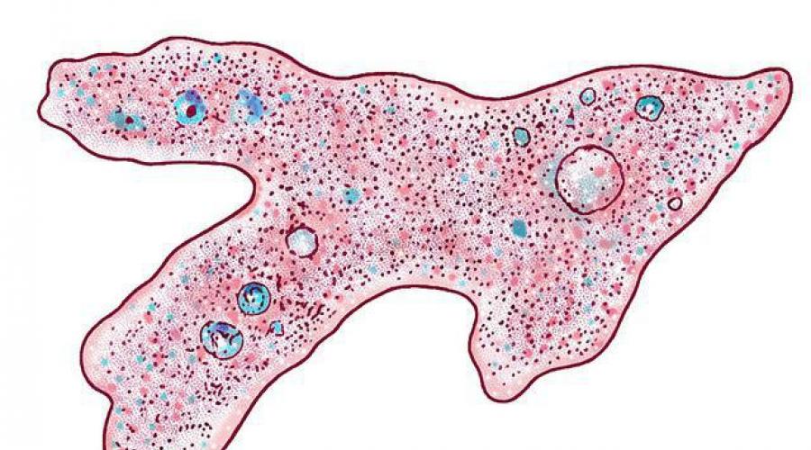
Amoeba is one of the most simply structured animals, devoid of a skeleton. It lives in the mud at the bottom of ditches and ponds. Externally, the body of the amoeba is a grayish gelatinous lump 200-700 microns in size, which does not have a permanent shape, which consists of cytoplasm and a vesicular nucleus and does not have a shell. Protoplasm contains an outer, more viscous (ectoplasm) and an inner granular, more liquid (endoplasm) layer.
On the body of the amoeba, outgrowths that change their shape are constantly formed - false legs (pseudopodia). Cytoplasm gradually flows into one of these protrusions, the false stalk attaches to the substrate at several points, and the amoeba moves. While moving, the amoeba encounters unicellular algae, bacteria, small unicellular organisms, and covers them with pseudopods so that they end up inside the body, forming a digestive vacuole around the swallowed piece in which intracellular digestion occurs. Undigested residues are thrown out in any part of the body. The method of capturing food using false legs is called phagocytosis. The liquid enters the amoeba’s body through the thin tube-like channels that are formed, i.e. by pinocytosis. End products of life ( carbon dioxide and other harmful substances and undigested food debris) are excreted with water through a pulsating (contractile) vacuole, which removes excess fluid every 1-5 minutes.
The amoeba does not have a special respiratory organelle. It absorbs the oxygen necessary for life over the entire surface of the body.
Amoebas reproduce only asexually (mitosis). IN unfavorable conditions(for example, when a reservoir dries out), amoebas retract pseudopodia, become covered with a durable double membrane and form cysts (encysts).

When exposed to external stimuli (light, changes in the chemical composition of the environment), the amoeba responds with a motor reaction (taxis), which, depending on the direction of movement, can be positive or negative.
Other class representatives
Many species of sarcodidae live in marine and fresh waters. Some sarcoids have a shell-shaped skeleton on the surface of the body (shell rhizomes, foraminifera). The shells of such sarcoids are permeated with pores, from which pseudopodia protrude. In shell rhizomes, reproduction is observed by multiple fission - schizogony. Marine rhizomes (foraminifera) are characterized by alternating asexual and sexual generations.
Possessing a skeleton, sarcodae are among the oldest inhabitants of the Earth. Chalk and limestone were formed from their skeletons. Each geological period is characterized by its own foraminifera, and the age of geological strata is often determined from them. The skeletons of certain types of shell rhizomes accompany the deposition of oil, which is taken into account during geological exploration.
Dysenteric amoeba(Entamoeba histolytica) is the causative agent of amoebic dysentery (amoebiasis). Discovered by F. A. Lesh in 1875

Localization. Human intestines.
. Everywhere, but more often in countries with hot climates.
Morphological features and life cycle. In the human intestine life cycle The following forms are found:
- cysts - 1, 2, 5-10 (Fig. 2).
- small vegetative form living in the intestinal lumen (forma minuta) - 3, 4;
- large vegetative form living in the intestinal lumen (forma magna) - 13-14
- tissue, pathogenic, large vegetative form (forma magna) - 12;
A characteristic feature of dysentery amoeba cysts is the presence of 4 nuclei in them (a distinctive feature of the species), the size of the cysts is from 8 to 18 microns.
Dysenteric amoeba usually enters the human intestine in the form of cysts. Here, the shell of the swallowed cyst dissolves and a quadruple amoeba emerges from it, which quickly divides into 4 single-nucleate small (7-15 microns in diameter) vegetative forms (f. minuta). This is the main form of existence of E. histolytica.
The small vegetative form lives in the lumen of the large intestine, feeds mainly on bacteria, reproduces and does not cause disease. If conditions are not favorable for the transition to the tissue form, then the amoebas, entering the lower intestines, encyst (turn into a cyst) with the formation of a 4-nuclear cyst and are excreted into the external environment with feces.
If conditions favor the transition to the tissue form (E. histolytica forma magna), the amoeba increases in size to an average of 23 microns, sometimes reaching 30 and even 50 microns, and acquires the ability to secrete hyaluronidase, proteolytic enzymes that dissolve tissue proteins and penetrate the walls intestines, where it multiplies intensively and causes damage to the mucous membrane with the formation of ulcers. In this case, the walls of blood vessels are destroyed and bleeding occurs into the intestinal cavity.
When amoebic intestinal lesions appear, small vegetative forms located in the intestinal lumen begin to transform into a large vegetative form. The latter is characterized by large size (30-40 microns) and the structure of the nucleus: the chromatin of the nucleus forms radial structures, a large lump of chromatin - a karyosome - is located strictly in the center, the forma magna begins to feed on red blood cells, i.e. it becomes an erythrophage. Characterized by blunt, wide pseudopodia and jerky movement.
Amoebas that multiply in the tissues of the intestinal wall - the tissue form - enter the intestinal lumen and become similar in structure and size to the large vegetative form, but are not able to swallow red blood cells.
With treatment or an increase in the body's protective reaction, the large vegetative form (E. histolytica forma magna) again turns into a small one (E. histolytica forma minuta), which begins to encyst. Subsequently, either recovery occurs, or the disease becomes chronic.
The conditions necessary for the transformation of some forms of dysenteric amoeba into others were studied by the Soviet protistologist V. Gnezdilov. It turned out that various unfavorable factors - hypothermia, overheating, malnutrition, overwork, etc. - contribute to the transition of forma minuta to forma magna. A necessary condition is also the presence of certain types of intestinal bacteria. Sometimes an infected person secretes cysts for many years without signs of disease. Such people are called cyst carriers. They pose a great danger as they serve as a source of infection for others. One cyst carrier releases up to 600 million cysts per day. Cyst carriers are subject to identification and mandatory treatment.
The only one source of the disease amoebiasis - man. Cysts released in feces contaminate soil and water. Since feces are often used as fertilizer, cysts end up in gardens and gardens, where they contaminate vegetables and fruits. Cysts are resistant to exposure external environment. They enter the intestines with unwashed vegetables and fruits, through unboiled water, dirty hands. Mechanical carriers are flies and cockroaches that contaminate food.
Pathogenic effect. When the amoeba penetrates the intestinal walls, a serious disease develops, the main symptoms of which are: bleeding ulcers in the intestines, frequent and loose stools (up to 10-20 times a day) mixed with blood and mucus. Sometimes, through the blood vessels, the dysenteric amoeba - erythrophage - can be carried into the liver and other organs, causing the formation of abscesses (focal suppuration) there. If left untreated, the mortality rate reaches 40%.
Laboratory diagnostics. Microscopy: fecal smears. In the acute period, the smear contains large vegetative forms containing red blood cells; cysts are usually absent, since f. magna is unable to encyst. In the chronic form or cyst carriage, quadruple cysts are found in the feces.
Prevention: personal - washing vegetables and fruits boiled water, drinking only boiled water, washing hands before eating, after visiting the toilet, etc.; public - combating soil and water contamination with feces, extermination of flies, sanitary education work, screening for cyst carriage of persons working in enterprises Catering, treatment of patients.
Non-pathogenic amoebae include intestinal and oral amoebae.

Intestinal amoeba (Entamoeba coli).
Localization. The upper part of the colon, lives only in the intestinal lumen.
Geographical distribution. Found in approximately 40-50% of the population various areas globe.
. Vegetative form has dimensions of 20-40 microns, but sometimes larger forms are also found. There is no sharp boundary between ectoplasm and endoplasm. Possesses in a characteristic way movement - simultaneously releases pseudopodia from different sides and, as it were, “marks time”. The nucleus contains large clumps of chromatin, the nucleolus lies eccentrically, radial structure absent. It does not secrete a proteolytic enzyme, does not penetrate the intestinal wall, and feeds on bacteria, fungi, and the remains of plant and animal food. The endoplasm contains many vacuoles. Does not swallow red blood cells, even if they are contained in the intestines in large quantities(in patients with bacterial dysentery). In the lower part of the digestive tract it forms eight- and two-core cysts.
Oral amoeba (Entamoeba gingivalis).
Localization. Oral cavity, dental plaque in healthy people and those with oral diseases, dental caries.
Geographical distribution. Everywhere.
Morphophysiological characteristics. The vegetative form has dimensions from 10 to 30 microns, highly vacuolated cytoplasm. The type of movement and structure of the nucleus resembles a dysentery amoeba. It does not swallow red blood cells; it feeds on bacteria and fungi. In addition, leukocyte nuclei or so-called salivary corpuscles are found in the vacuoles, which, after staining, may resemble red blood cells. It is believed that it does not form cysts. The pathogenic effect is currently denied. It is found in dental plaque of healthy people in 60-70%. It is more common in people with dental and oral diseases.
Habitat "Common Amoeba"
The common amoeba is found in the sludge at the bottom of ponds with polluted water. It looks like a small (0.2-0.5 mm), barely visible to the naked eye, colorless gelatinous lump, constantly changing its shape (“amoeba” means “changeable”). The details of the amoeba's structure can only be seen under a microscope.
Structure and movement of the "common amoeba"
The body of the amoeba consists of semi-liquid cytoplasm with a small vesicular nucleus enclosed inside it. An amoeba consists of one cell, but this cell is a whole organism leading an independent existence.
The cytoplasm of the cell is in constant movement. If the current of cytoplasm rushes to one point on the surface of the amoeba, a protrusion appears in this place on its body. It enlarges, becomes an outgrowth of the body - a pseudopod, cytoplasm flows into it, and the amoeba moves in this way. Amoeba and other protozoa capable of forming pseudopods are classified as rhizopods. They received this name due to the external resemblance of their pseudopods to plant roots.
Food "Ameba vulgaris"
An amoeba can simultaneously form several pseudopods, and then they surround food - bacteria, algae, and other protozoa. From the cytoplasm surrounding the prey, digestive juice is secreted. A bubble is formed - a digestive vacuole.
Digestive juice dissolves some of the substances that make up food and digests them. As a result of digestion, they form nutrients, which leak from the vacuole into the cytoplasm and go to build the body of the amoeba. Undissolved residues are thrown out anywhere in the amoeba’s body.
Breathing "Ameba vulgaris"
The amoeba breathes oxygen dissolved in water, which penetrates its cytoplasm through the entire surface of the body. With the participation of oxygen, complex food substances in the cytoplasm are decomposed into simpler ones. This releases energy necessary for the functioning of the body.
Release of harmful substances from vital activity and excess water "Vulgar Amoeba"
Harmful substances are removed from the amoeba’s body through the surface of its body, as well as through a special vesicle - contractile vacuole. The water surrounding the amoeba constantly penetrates the cytoplasm, diluting it. The excess of this water from harmful substances gradually fills the vacuole. From time to time, the contents of the vacuole are thrown out.
So, food, water, and oxygen enter the amoeba’s body from the environment. As a result of the life activity of the amoeba, they undergo changes. Digested food serves as material for building the body of the amoeba. Substances that are harmful to the amoeba are removed outside. Happening metabolism of amoeba vulgaris. Not only amoeba, but also all other living organisms cannot exist without metabolism both within their body and with the environment.
Reproduction of "Ameba vulgaris"
 The amoeba's nutrition causes its body to grow. The grown amoeba begins to reproduce. Reproduction begins with a change in the nucleus. It stretches out, is divided by a transverse groove into two halves, which diverge into different sides- two new nuclei are formed. The body of the amoeba is divided into two parts by a constriction. Each of them contains one core. The cytoplasm between both parts is torn and two new amoebas are formed. The contractile vacuole remains in one of them, but appears anew in the other. So, the amoeba reproduces by dividing in two. During the day, division can be repeated several times.
Cyst
 Amoeba feeds and reproduces throughout the summer. In autumn, when cold weather sets in, the amoeba stops feeding, its body becomes rounded, and a dense protective shell is formed on its surface - a cyst is formed. The same thing happens when the pond where the amoebas live dries out. In the state of a cyst, the amoeba tolerates unfavorable living conditions. When favorable conditions occur, the amoeba leaves the cyst shell. She releases pseudopods, begins to feed and reproduce. Cysts carried by the wind contribute to the spread of amoebas.
Amoebas are a detachment of the smallest single-celled organisms from the subclass of rhizomes of the class Sarcodae, such as sarcomastigophores. Distinctive feature All representatives of this group of protozoa have the ability to form pseudopods (pseudopodia) for movement and capture of food. Pseudopodia are outgrowths of the cytoplasm, the shape of which is constantly changing.
Amoeba is considered one of the simplest forms of life. However, from a physiological point of view, an amoeba cell is quite complex arranged system. In the body of the amoeba, functions characteristic of higher multicellular organisms are carried out - respiration, excretion, digestion.
All amoebas have irregular shape, which is constantly changing due to the formation of pseudopods. This adaptation, as mentioned above, was formed in the process of evolution for nutrition and movement. These organisms lack a dense membrane around the cell. There is only a special molecular layer called the plasma membrane, which is an integral element of the living cytoplasm.
The internal structure of the amoeba has characteristics. The cytoplasm is divided into inner part(endoplasm) and external (ectoplasm). Endoplasm has a granular structure, and ectoplasm has an approximately uniform consistency. The endoplasm contains a large nucleus, contractile and digestive vacuoles, and fatty inclusions.
Organisms in this group feed on protozoa, bacteria, and algae. With the help of pseudopodia, food is captured by the amoeba and enters its endoplasm, where a digestive vacuole is formed in which food particles are digested. The release of undigested residues, as well as waste products, occurs in amoebas through the entire surface of the body through ordinary diffusion.
The function of the contractile vacuole is to remove excess water from the body of the individual. When the vacuole contracts, it pushes water out.
Amoebas reproduce asexually by binary fission. A constriction forms in the mother cell, and the cytoplasm is divided into two approximately equal parts with a nucleus in each. The nuclei of young individuals are formed as a result of mitotic division of the nucleus of the mother cell. Two young amoebas gradually grow and at a certain stage divide again, giving rise to new individuals.
The common amoeba is a cell in appearance and is directly related to the type of protozoa, to the class of rhizomes, or they are also called Sarcodaceae. They have pseudopods, which are organs with which they move and capture food. The cell does not have a dense membrane, and therefore the amoeba can easily change its shape. The outer covering is a very thin cytoplasmic membrane.
Amoeba ordinary structure.
Amoeba has a very simple structure. One of the simplest living creatures. Has no skeleton. The common amoeba lives at the bottom of various reservoirs, in silt. There is one thing: in bodies of water only fresh water: a pond, a ditch, etc. If you look at it, you will notice that this gray transparent lump does not have a permanent shape. The name of this creature translates as “changeable.” Pseudopods are constantly forming on the cell body, due to the fact that the cytoplasm flows back and forth. The size of the lump can be at least 0.2 millimeters and, at most, 0.7 millimeters. Organelles - pseudopods contribute to the movement of this tiny creature. The movement is very slow, it resembles the flow of thick mucus. During its movement, the amoeba encounters various single-celled organisms, such as algae and bacteria. It flows around them and, as it were, absorbs them with its own cytoplasm, and a digestive vacuole is formed.
The common amoeba secretes specific enzymes in its cytoplasm that digest food. The process of intracellular digestion occurs. Digested foods in liquid form enter the cytoplasm itself, and undigested food remains are thrown away. This method of food capture is called phagocytosis. The body of the amoeba has thin channels through which fluid enters the cell body. This process is called pinocytosis. There is one vacuole that throws excess liquid products out. It's called Eliminate excess every five minutes. The endoplasm contains a nucleus. Reproduction occurs as follows: the cell divides in half, that is, asexually.
How an amoeba protects itself from adverse external influences.
Common amoeba and dysenteric amoeba are Move with the help of organelles-psepododes, belong to the rhizopods;
The class of rhizomes resembles algae, which indicates their relationship;
It feeds on what it gets from other plants, or from others, which is what distinguishes amoebas from algae.
Amoeba is, although the simplest, a whole organism capable of leading an independent existence.